
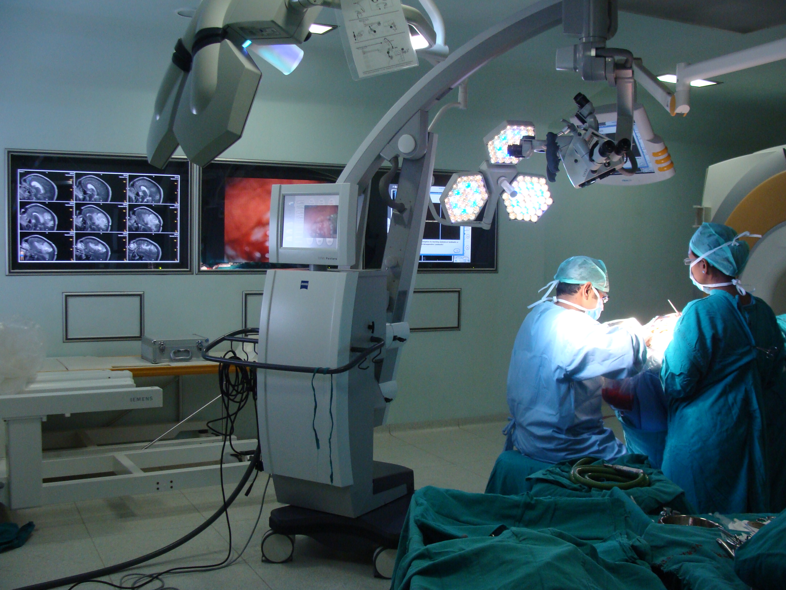
Localizing the epileptogenic foci using the imaging data: Establish the imaging and electrical localization for the ‘epileptogenic focus’ in patients being investigated for epilepsy surgery. This will be first achieved by the use of the existing armamentarium at AIIMS like EEG, VEEG, MRI, SPECT, and PET. To this the information from Magnetoencephalography (MEG) facility will be added. These imaging information will then be co-registered on the actual patient using a special fusion software downloaded on to an advanced neuro-navigation system.
Objective intraoperative electrocorticography: During the resective surgery, first an intraoperative electrocorticography (ECoG) will be performed to mark out in an objective manner (using a quantitative scoring system) the different degree of abnormalities of electrical activity recorded from the surface of the brain. Following this, different brain samples will be collected within the resective surgery zone and will be processed for further cytological and molecular studies. Thus, every portion of brain sample removed for processing will have a quantifiable imaging score, MEG score and an ECoG score.